Advanced Technology in Thornhill
When you visit your eye doctor, you want to know that the optometry office has the best and most advanced technology to diagnose eye disease, and screen for possible vision problems. At Disera Optical we use the most advanced technology in all of our eye exams and eye care services. Read about the different technologies we use during your complete eye exam.
Corneal Mapping
Corneal topography, also known as photokeratoscopy or videokeratography, is a non-invasive medical imaging technique for mapping the surface curvature of the cornea, the outer structure of the eye. Since the cornea is normally responsible for some 70% of the eye's refractive power, its topography is of critical importance in determining the quality of vision.
The three-dimensional map is therefore a valuable aid to the examining ophthalmologist or optometrist and can assist in the diagnosis and treatment of a number of conditions; in planning refractive surgery such as LASIK and evaluation of its results; or in assessing the fit of contact lenses. A development of keratoscopy, corneal topography extends the measurement range from the four points a few millimeters apart that is offered by keratometry to a grid of thousands of points covering the entire cornea. The procedure is carried out in seconds and is completely painless.
Special thanks to the EyeGlass Guide, for informational material that aided in the creation of this website. Visit the EyeGlass Guide today!
Digital Retinal Imaging
We use cutting-edge digital imaging technology to assess your eyes. Many eye diseases, if detected at an early stage, can be treated successfully without total loss of vision. Your retinal Images will be stored electronically. This gives the eye doctor a permanent record of the condition and state of your retina.
This is very important in assisting your Optometrist to detect and measure any changes to your retina each time you get your eyes examined, as many eye conditions, such as glaucoma, diabetic retinopathy and macular degeneration are diagnosed by detecting changes over time.
The advantages of digital imaging include:
- Quick, safe, non-invasive and painless
- Provides detailed images of your retina and sub-surface of your eyes
- Provides instant, direct imaging of the form and structure of eye tissue
- Image resolution is extremely high quality
- Uses eye-safe near-infra-red light
- No patient prep required
Digital Retinal Imaging
Digital Retinal Imaging allows your eye doctor to evaluate the health of the back of your eye, the retina. It is critical to confirm the health of the retina, optic nerve and other retinal structures. The digital camera snaps a high-resolution digital picture of your retina. This picture clearly shows the health of your eyes and is used as a baseline to track any changes in your eyes in future eye examinations.
Visual Field Testing
A visual field test measures the range of your peripheral or “side” vision to assess whether you have any blind spots (scotomas), peripheral vision loss or visual field abnormalities. It is a straightforward and painless test that does not involve eye drops but does involve the patient's ability to understand and follow instructions.
An initial visual field screening can be carried out by the optometrist by asking you to keep your gaze fixed on a central object, covering one eye and having you describe what you see at the periphery of your field of view. For a more comprehensive assessment, special equipment might be used to test your visual field. In one such test, you place your chin on a chin rest and look ahead.
Lights are flashed on, and you have to press a button whenever you see the light. The lights are bright or dim at different stages of the test. Some of the flashes are purely to check you are concentrating. Each eye is tested separately, and the entire test takes 15–45 minutes. These machines can create a computerized map of your visual field to identify if and where you have any deficiencies, which can be important when evaluating vision suitability for treatments such as Orthokeratology CRT & VST.
Advanced Techonology Used at Disera Optical
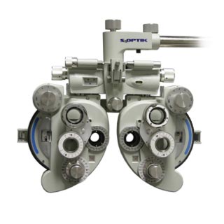
S4OPTIK SLY-100 REFRACTOR
Humphrey Matrix 800
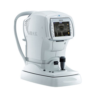
Righton Speedy-K2
NT-510 Non-Contact Tonometer

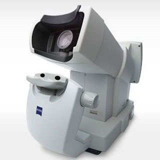
AFC-330 Fundus Camera
Thornhill, ON L4J 0A7
- Monday: 10:00 AM - 6:00 PM
- Tuesday: 10:00 AM - 6:00 PM
- Wednesday: 10:00 AM - 6:00 PM
- Thursday: 10:00 AM - 6:00 PM
- Friday: 10:00 AM - 6:00 PM
- Saturday: 9:00 AM - 2:00 PM
- Sunday: Closed